The things and objects of history are important because they provide a tangible connection to the past. Seeing, or better yet holding and touching, the stuff that generations now dead made and worked with enlivens history, shucking us from the present and its endless clamour for our attention.
The Hidden Structures exhibition at the Science Museum trips us into the history of X-ray crystallography with a small but intriguing display of objects from the 1940s through to the 1970s. The exhibition commemorates the centenary of the development of the technique, by the father and son team of William and Lawrence Bragg who figured out how the scattering of X-rays by crystals could be analysed to reveal the atomic and molecular arrangements within, providing a vista of the structure of matter that had never been seen before.
The Braggs first applied the technique in 1913 to show how the patterns of X-rays diffracted onto photographic plates by table salt — sodium chloride — could be interpreted to reveal the organisation of the two atoms within its crystals. It was apparent from the beginning that the method was applicable to anything that could be induced to crystallise, even the most complex molecules of chemistry and biology. Soon structures composed of tens or hundreds or even thousands of atoms were emerging from UK labs, which established itself at the forefront of the technique thanks in no small part to the inspirational leadership of the younger Bragg.
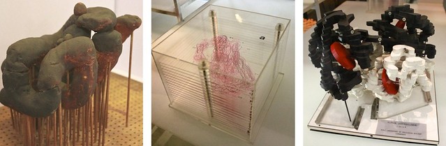
The first protein structures; left to right — myoglobin, perspex stack of electron density, haemoglobin
The artefacts in the Hidden Structures display come mainly from the first bloom of chemical and biological crystallography; there is Dorothy Hodgkin’s ball-and-stick model of penicillin, John Kendrew’s wormy brown representation of the oxygen-storage protein, myoglobin, Max Perutz’s black and white slabbed structure of haemoglobin, the oxygen-transporter from human blood, and in pride of place, Hodgkin’s huge model of the atomic structure of insulin.
These scientists had to be very hands-on at all stages of their work — growing crystals, carefully measuring X-ray diffraction patterns recorded on photographs, and printing out the electron density maps produced by their analysis. These three-dimensional maps (there is one for haemoglobin in the display, printed in sections on stacked sheets of perspex) show where the electrons are concentrated, so defining the positions of the atoms. The early models simply depict the contours of these maps and give the overall form of the protein molecule. Coming some years after X-rays had unveiled the elegant double-helix of DNA, their crude irregularity was at first a disappointment: “hideous and visceral” wrote Perutz.
But the resolving power of X-rays soon improved and those early crystallographers had to swap plastic and plasticene for intricate assemblies of rods, each representing a bond between two atoms, that were put together with loving attention to detail. Hodgkin’s insulin model from the early 1960s may not be beautiful, but it is mesmerising — and hugely informative.
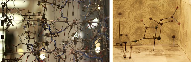
Hodgkin’s atomic structures: left, insulin; right, penicillin.
X-ray crystallography continues apace. Thousands of protein structures have been solved, providing a detailed understanding of the workings of biology at the molecular level. We see clearly now not just how hormones like insulin work, or how haemoglobin picks up and drops off its cargo of oxygen, but also how DNA is synthesised and decoded, how ion channels enable the transmission of nerve signals, how the immune system fights off infection. No pharmaceutical company works blind in the 21st Century; all use X-ray crystallography to reveal the molecular targets of therapy, whether from a virus or bacteria or a cancerous cell, as part of the quest for new drugs and vaccines.
But all the work of recording and analysing data and building models has now migrated to computers. For sure this has greatly accelerated the pace of research and discovery, but there are no more photographs or stacks of electron density or models made of stuff for future generations to pick up and wonder at. All the more reason therefore to cherish the crystallographic arcana on show at the Science Museum.
Stephen Curry is a professor of structural biology at Imperial College. He writes regularly about science at the Reciprocal Space and Occam’s Corner blogs.