When physics – in the form of X-ray diffraction – collided with biology in the middle of the last century, scientists were given an intimate glimpse of the vast, complex molecules that perform the fundamental processes of life.
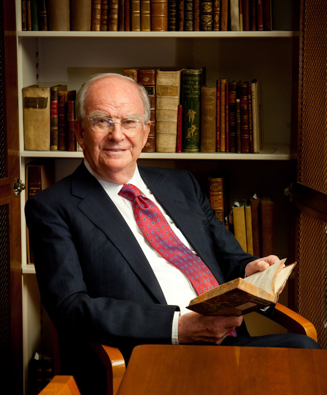
To mark the publication of a new book about the British pioneers of the field, the Science Museum hosted an event this week with Sir John Meurig Thomas, a wizard when it comes to harnessing matter in the form of catalysts to eliminate the solvents and manufacturing steps of industrial processes, making them greener.
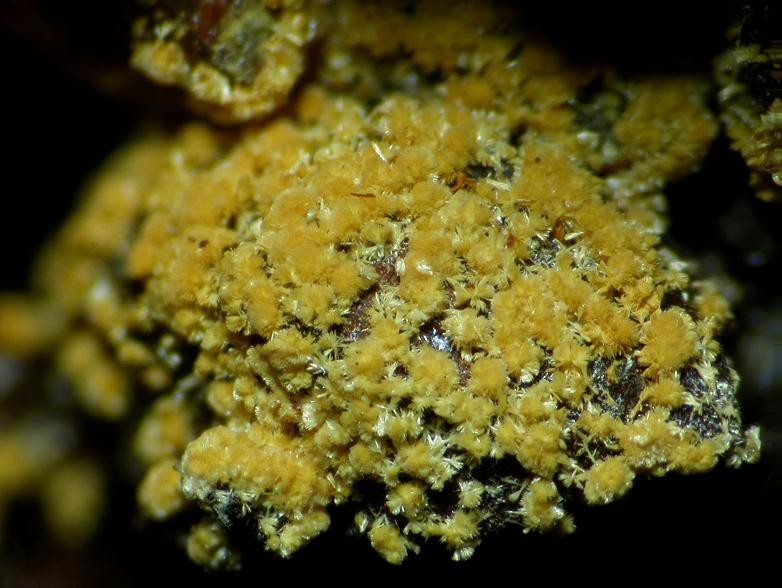
Sir John’s distinguished career has seen him earn 23 Honorary Doctorates notably for his work on catalysts, such as zeolites and clays, where important chemical reactions could be speeded up inside their molecule-sized pores, chambers and cavities.
Interviewing Sir John was Science Museum Group’s Chair, Dame Mary Archer. Both are chemists; both overlapped at the University of Cambridge, when John was the Head of the Department of Physical Chemistry in the late 1970s-80s, and both have links to the Royal Institution, where Dame Mary did postdoctoral research and Sir John was later Director.
That is one of the reasons why, in 1991, Sir John was knighted “for services to chemistry and the popularisation of science”.
Sir John’s new book, Architects of Structural Biology, published by Oxford University Press, celebrates the 20th century fusion of chemistry and physics, which furnished a way to reveal the atomic structure of complex molecules with X-rays, using a method called X-ray diffraction.
In his book he focuses on four individuals – all of whom Sir John knew personally – who cleared the way for the routine use of X-ray diffraction, transforming medicine and the biosciences.
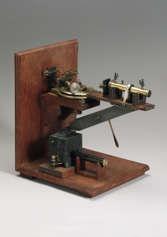
There was Lawrence Bragg (1890–1971), who first developed X-ray diffraction, where the scattering of X-rays (which can’t be focused) by a crystal are interpreted in terms of its atomic symmetries and structure. To do this, he devised an instrument that could take photographs of the spotty patterns made by scattered X-rays, called an X-ray spectrometer and the original (1910-1926) can be seen on display in the Science Museum’s Making the Modern World Gallery.
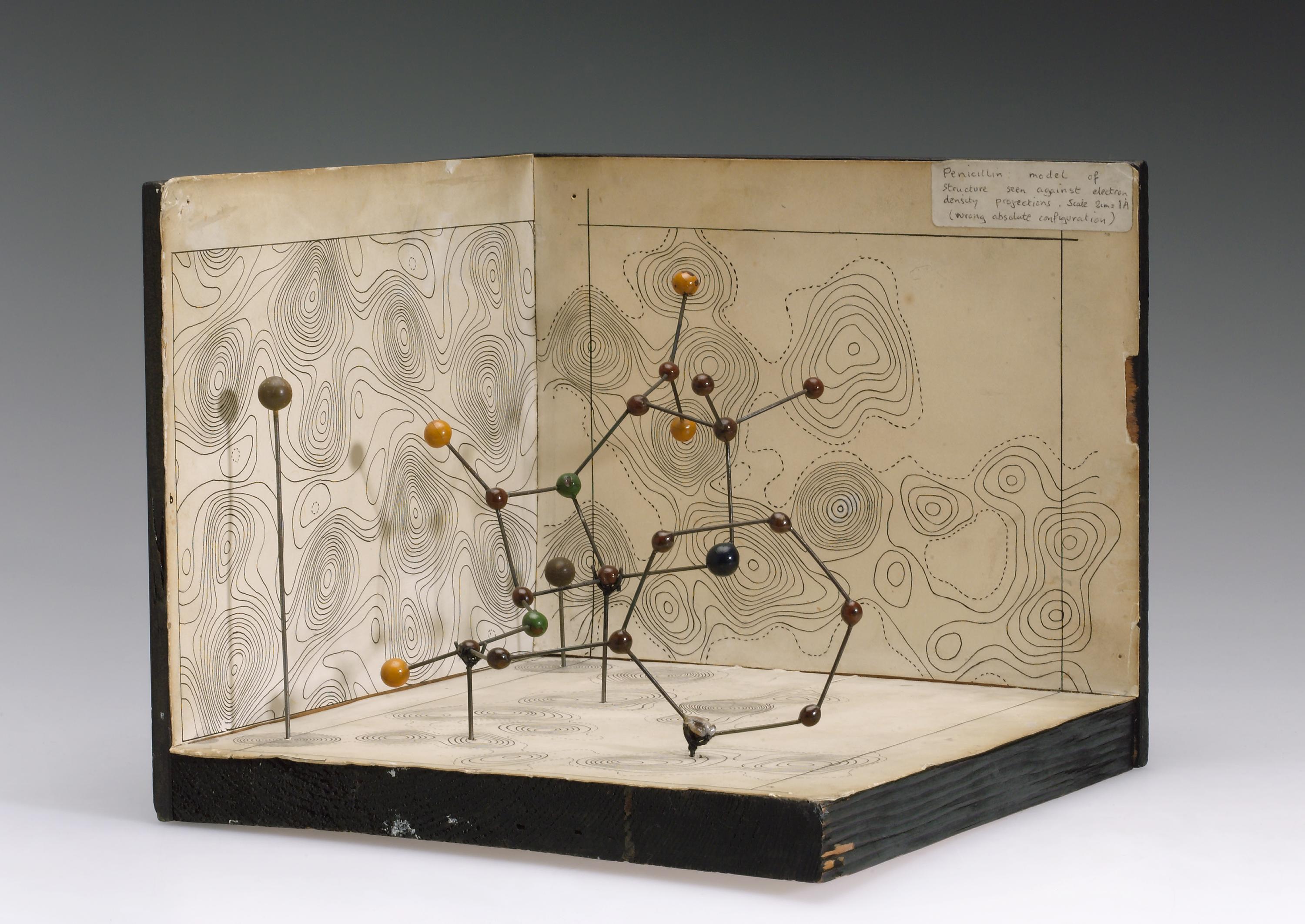
The other three pioneers in his book — Max Perutz (1914-2002), John Kendrew (1917-1997), and Dorothy Hodgkin (née Crowfoot, 1910-1994) – put this technique to work in biology.
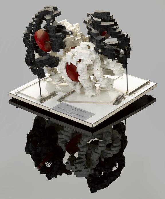
In our collections, you can see Hodgkin’s molecular models of penicillin and insulin, while on display in Medicine: The Wellcome Galleries, stands Kendrew’s model of the muscle protein myoglobin, where the supporting rods are easier to see than the model itself, and nearby is Francis Crick and James Watson‘s molecular model of the DNA double helix, which revealed how heredity is passed down the generations. All these structures were determined by X-ray crystallography.
With these molecular models came understanding of how they worked. Take the example of haemoglobin, the red pigment in blood. In 1959, Max Perutz determined its molecular structure by X-ray crystallography, revealing it contains four atoms of iron, each held at the heart of an array of organic rings called a porphyrin, responsible for the red colour. That arrangement allows oxygen to ‘look but not touch’ and bind reversibly, so rust does not result.
Once you know the structure, you can understand how a genetic mutation in the gene for haemoglobin causes sickle cell disease, or why carbon monoxide is lethal.
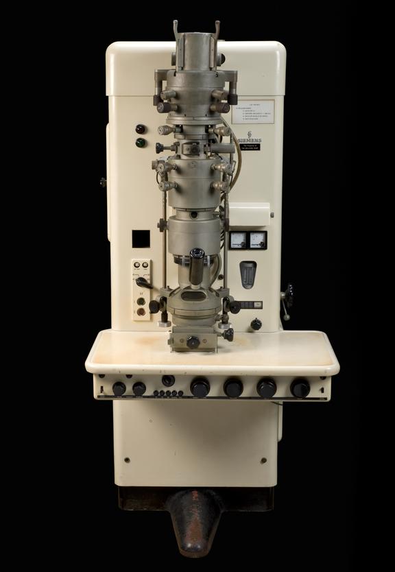
Dame Mary and Sir John also discussed how the technique of X-ray diffraction has in part been overtaken by an extension of electron microscopy, called cryo-EM, which is now reaching atomic resolution, though Sir John believes there will still be heavy demand for conventional X-ray diffraction from pharmaceutical companies.
Cryo-EM was pioneered by Richard Henderson at the MRC’s Laboratory of Molecular Biology, LMB, in Cambridge, an institution that has garnered many Nobel prizes for uncovering the chemical structures of life, including one for Henderson himself.
In their discussion, Dame Mary and Sir John ranged from Dorothy Hodgkin’s inspirational story and the creative culture of the LMB to his unease about the effects of Brexit on international collaborations, such as work on synchrotrons, intense sources of X-rays, and at the Institut Laue-Langevin, a research reactor in Grenoble that is used as a powerful source of neutrons.
They also touched on Sir John’s extraordinary career which had unpromising beginnings near Llanelli, South Wales, where his father and brother were miners.
The options at that time were ‘to become a preacher, teacher or work underground,’ Sir John recalled. His brother was 12 years his senior, and he reflected that if their births had been reversed, ‘I would have ended up a coal miner and he a professor. He was a very intelligent man, as was my father.’
Sir John’s interest in science was kindled as a teenager by his physics teacher at Gwendraeth Grammar School. ‘She was astonishingly good, talking about the great scientists such as Newton and Rutherford and at length about Michael Faraday. I was enthralled when I heard her describe how this man who had left school at the age of 13 with little knowledge, other than arithmetic, became a bookbinder’s assistant and then ended up being one of the greatest scientists of all time. I was in awe of Faraday from that moment onward.’
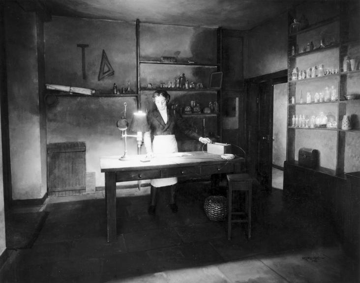
Later in life, Sir John would become the Fullerian Professor of Chemistry at the Royal Institution, London and then the Director, positions held by his hero. Sir John lived in the same living quarters that Faraday and his wife once occupied in the Royal Institution’s building on Albemarle Street.
During his inaugural lecture, sitting before him was Irene James, the brilliant and well-read schoolmistress who had first fired up his interest in science. ‘It was a great moment.’
Sir John earned his BSc in 1954 and doctorate a few years later from the University of Wales, Swansea, doing ‘quite well.’
After spending a year working at the United Kingdom Atomic Energy Authority as scientific officer, where he ‘rubbed shoulders with metallurgists who spoke a language I did not understand’, when it came to defects, dislocations and imperfections, and that inspired him to take an interest in their consequences for chemistry rather than the strength of materials.
He joined the Department of Chemistry at the University of Wales, Bangor where he rose through the ranks from Assistant Lecturer to Reader, and did research to reveal the profound influence that structural imperfections such as dislocations have on the chemical properties of solids.
While there he pioneered the use of electron microscopy – what he describes as his ‘single best’ decision in his scientific research. ‘I realise that if I wanted to study the chemistry of defects and imperfections in solids, and catalytic and reactivity, I needed an electron microscope.’
In 1959, Sir John married Margaret Edwards with whom he later had two daughters, Lisa and Naomi.
In 1969 he became Professor and Head of Chemistry at the University College of Wales, Aberystwyth, where he broadened his interests in solid-state chemistry to create ‘ one of the finest Departments of solid state chemistry in Europe. That’s where I did a good deal of mineralogy also. And that is where my work on defects attracted the attention of Cambridge.’
It also caught the attention of the prestigious Royal Society. In 1977 he was elected a Fellow and, the following year, he joined the University of Cambridge as Head of the Department of Physical Chemistry, where he overlapped with Dame Mary in the late 70s/80s and continued working on new techniques such as neutron scattering (on which I did my own research) along with nuclear magnetic resonance, to study zeolites.
In 1986, BP used one of his patented acid catalysts to make hundreds of thousands of tons of ethyl acetate – in a single step – within the biggest plant of its kind in the world.
The same year, he was invited to succeed Nobelist Sir George Porter as Director of the Royal Institution. At this time, Sir John began using synchrotron radiation and devised techniques which combine X-ray spectroscopy and high-resolution X-ray diffraction to determine the atomic structure of the active sites of solid catalysts as they worked.
He also devised new mesoporous, microporous, and molecular sieve catalysts, and ‘single site’ heterogeneous catalysts, which have uniform sites where the catalysis takes place.
In 1987, the BBC televised his Royal Institution Christmas Lectures on crystals, with David Phillips, continuing the tradition of lectures for children started by Faraday in 1826.
Sir John resigned as Director in 1991, due to his wife’s poor health, but remained associated with the Davy Faraday Research Laboratory of the Royal Institution until 2006. In 1991 he published the book Michael Faraday and the Royal Institution: The Genius of Man and Place.

After a time as Deputy Pro-Chancellor of the University of Wales (1991–1994), Sir John returned to Cambridge in 1993 as Master of Peterhouse, the university’s oldest college, until 2002, the year his wife, Margaret, died. In April 2010 Sir John married Jehane Ragai of the American University in Cairo.
Sir John is the author of around 30 patents, holds honorary positions in several universities, is the recipient of 23 honorary degrees, has been elected to honorary membership in over 15 foreign academies, and has garnered a range of awards. Of all those, ‘one of the nicest’ came in 1995, when Sir John became the first British scientist in 80 years to be awarded the Willard Gibbs Award by the American Chemical Society.
The mineral meurigite is named after him and when Sir John celebrated his 75th birthday with a series of events at Cambridge, they were attended by Nobel laureates and none other than the world’s best-known quantum chemist, Angela Merkel, whom he knew through her husband, computational chemist Joachim Sauer, with whom he had worked while at the Royal Institution.
Among the audience was Lord Rees, Astronomer Royal, and Sir Venki Ramakrishnan, Nobel laureate and President of the Royal Society In the foreword to Sir John’s book, Sir Venki writes that the term ‘molecular biology’ has now been co-opted to mean the study of information and control in living systems, and that Sir John’s book restores the term to its original meaning: ‘After all, the remarkable advances in our understanding of genetics and control would not have been possible without the structure of DNA, which, in turn, would not have been possible without decades of study of the structure of molecules using X-ray diffraction.’