As one of the conservators preparing objects for display in the new Medicine Galleries, due to open in 2019, I have been fortunate to work on a wide variety of objects ranging from a 19th century post mortem set to a 21st century DNA analyser.
I love working on such an interesting collection and there is always some excitement when a new object is brought into the lab. I recently had the opportunity to work on one of Dr. Louis Auzoux’s anatomical models.
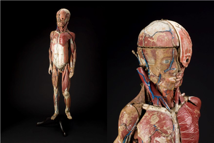
Dr. Auzoux was a French doctor, who in the 1820s developed a method of producing anatomical models made from papier-mâché that could be taken apart to instruct medical students about anatomy.
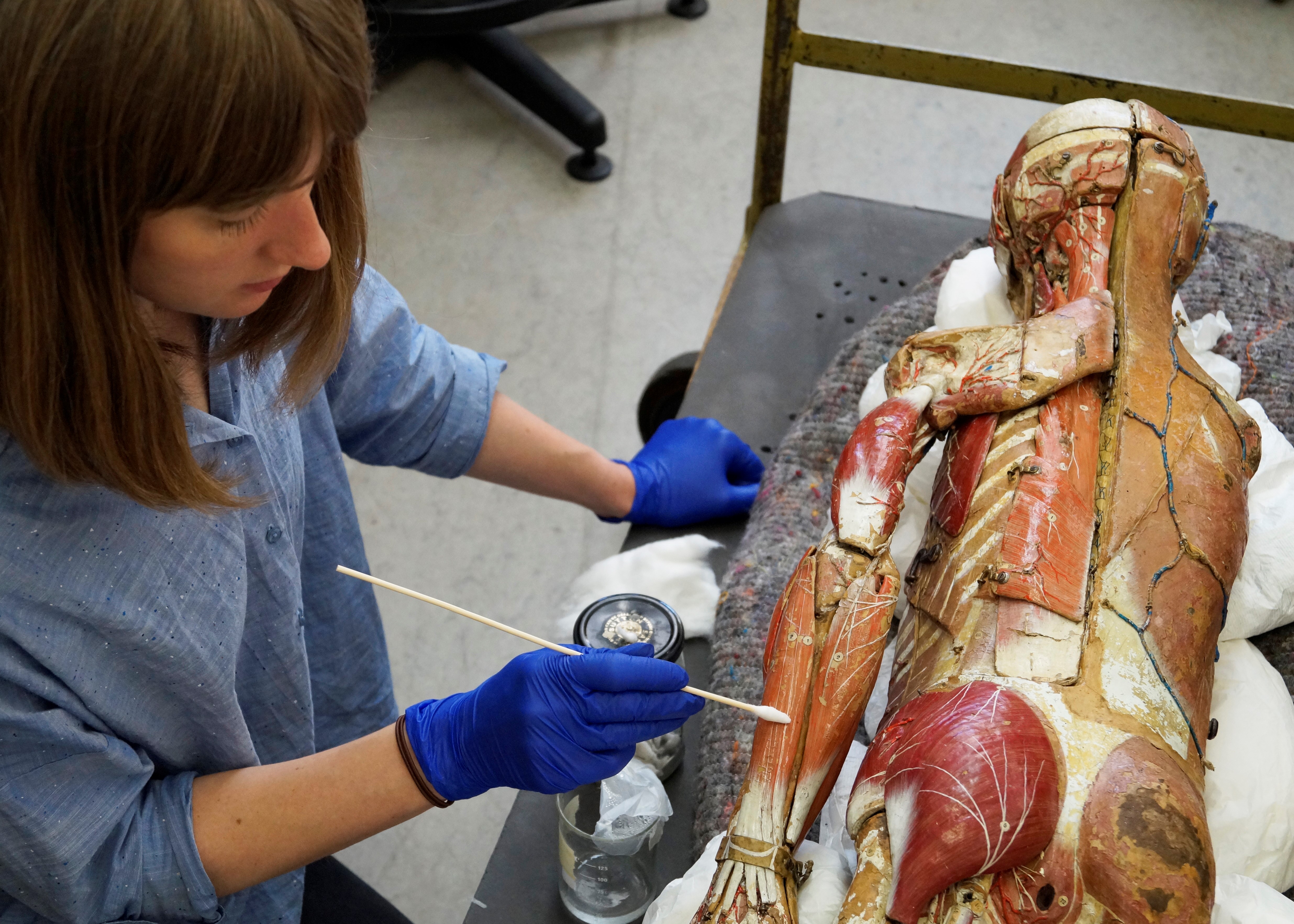
The object I worked on was a model of a male figure that was made in the workshops of Dr Auzoux between 1833 and 1866. It was a privilege to handle this object and remove some of the parts to discover the internal structure of the model. Not only was it a fascinating object but also one of my bigger conservation challenges.
I needed to document, clean, repair and stabilise the model so that it can be displayed and enjoyed by visitors in the new Medicine galleries.
There were numerous areas of flaking paint and loss to the paint layers, so my priority was to stabilise the flaking paint to prevent further loss. The model has a top layer of gelatine varnish, so I re-activated the gelatine using a heated spatula and adhered it back into position. A small amount of adhesive was used where this didn’t work.
Where I could remove some of the detachable muscles, I found areas where the papier-mâché layers had started to delaminate causing the muscles to deform. I used humidity to gently ease the layers back together, then adhesive and Japanese paper fills to stabilise the parts so that they could be safely re-attached to the model.
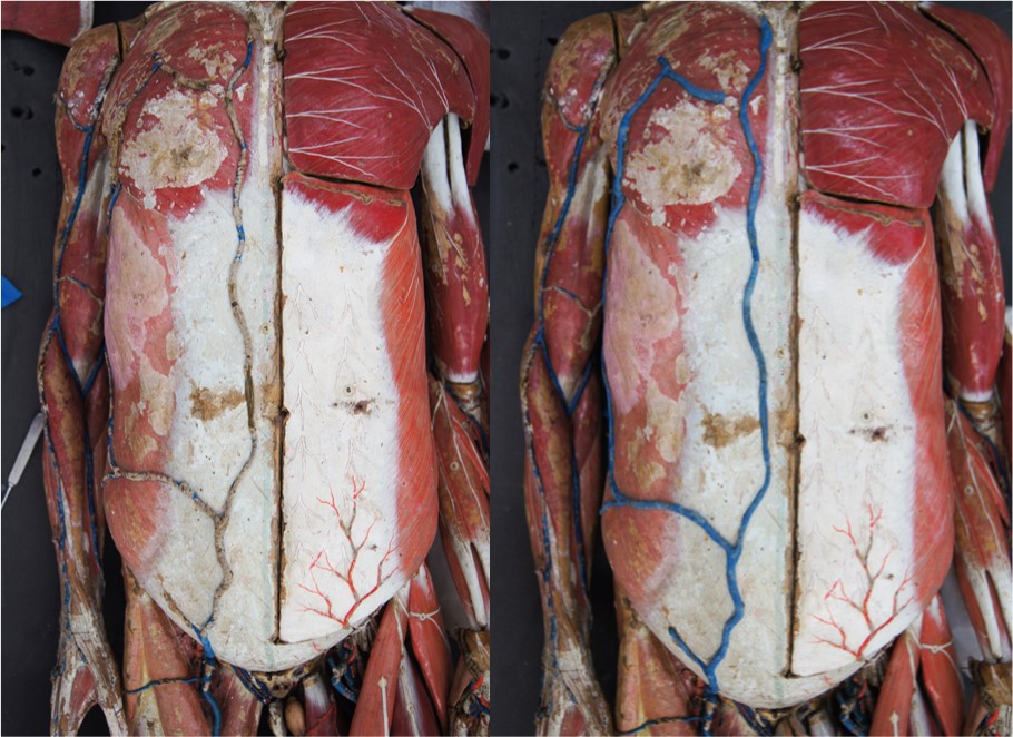
After discussion with the lead conservator and curator, I had the rare opportunity to carry out some restoration work on the model. There are veins running up the length of the model which show the flow of oxygen around the body. Unfortunately, the veins on the chest area had lost much of the blue paint, so their purpose was unclear. To restore these areas, I dyed Japanese tissue paper to match the original colour of the veins, then carefully overlaid and adhered thin strips of the tissue onto the raised veins.
After 35 hours of conservation treatment the model is now ready for display. Working on the model has been both enjoyable and beneficial in developing my conservation skills. I look forward to seeing the model on display and hope that visitors to the gallery will enjoy it for years to come.