Medicine is full of breakthroughs – and behind each one is a human story. Many of these groundbreaking innovations can be found on display within Medicine: The Wellcome Galleries, five vast and free galleries on the first floor of the Science Museum.
Stop 1: Omniskop X-ray apparatus, c.1925-1935
Start your tour by heading up to Medicine: The Wellcome Galleries via Lift or Stairs C. Enter the gallery and turn immediately right to find this large x-ray machine on display in the Imaging the Body section of the gallery.
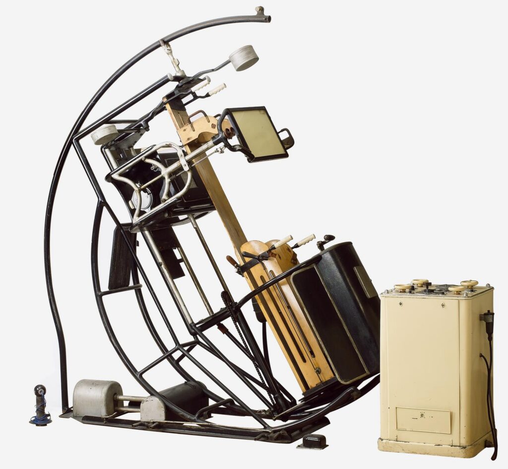
X-rays were the first imaging technology that revolutionised how the inside of living bodies could be seen and diagnosed.
This X-ray machine is called an ‘Omniskop’. It belonged to Ernst Rachwalsky (1889-1962), a German-Jewish doctor working in Berlin during the 1920s, who specialised in the treatment of stomach problems.
At the cutting-edge of x-ray technology at the time, the machine’s motor and counterweights meant that Dr Rachwalsky could tilt and position patients at different angles to help image and diagnose their conditions.
In the 1930s, as the Nazi regime tightened its grip, life became increasingly dangerous for Jewish people like Rachwalsky and his family. They decided to leave Germany and travel to England. Rather than leaving his machine behind, Rachwalsky shipped the Omniskop over in parts to London.
Setting up a new life as a doctor wasn’t easy. After having to take British medical qualifications and being detained for a time as an ‘enemy alien’ during the outbreak of the Second World War, Rachwalsky eventually set up a new medical practice on Wimpole Street in London, where he used the machine to diagnose patients – many of whom were also German-Jewish refugees – until his retirement into the 1950s.
In its own retirement, this Omniskop has been the centrepiece of exhibitions and has even inspired Hollywood movie set designers.
Stop 2: PillCam – A camera you can swallow
Find a display by the wall called Medicine by Looking. Look to the left of the showcase to find the PillCam.
What if diagnosing disease was as simple as taking a photo from the inside?
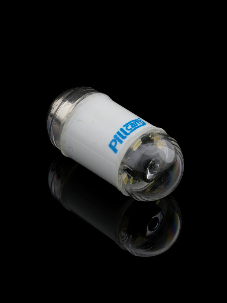
In 1999, Dr Paul Swain was the first person to swallow a PillCam – a tiny wireless camera designed to transmit thousands of pictures as it passes through the digestive system.
Watching the images live was Gavriel Iddan (1941-to date) an Israeli-Jewish engineer, who had spent over twenty years developing the idea of a swallowable camera many thought impossible.
Iddan came up with the idea of splitting the system into three parts: the miniature camera and its transmitter; a recorder worn by the patient; and software to process the data, enabling it to be reviewed later by a doctor, and explored the use of new types of silicon chips to make the camera small enough to swallow.
During the capsule’s 8 hour journey through the gut, the PillCam captures over 50,000 images, revealing parts of the intestines that were once nearly impossible to see without invasive procedures, making it a more patient-friendly way to detect signs of disease.
Iddan and Swain’s invention was compared to the miniaturised submarine that travels around the body in the 1966 science fiction film, Fantastic Voyage.
Stop 3: Rosalind Franklin and the discovery of DNA’s double helix structure
Walk towards the end of the gallery, until on the right-hand side you see this model of DNA. Look for the images of Rosalind Franklin and Photo 51 on the label strip.
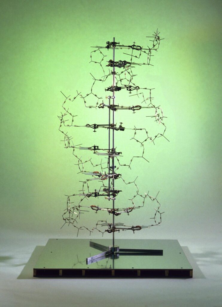
X-rays have proved useful not only for diagnosis, but also for revealing the structures of the most important molecules for life.
Born into a British Jewish family in London, Rosalind Franklin (1920–1958) was a brilliant chemist whose x-ray crystallography images of DNA made the discovery of its double-helix structure possible. She also worked on revealing the structures of viruses.
In 1952, Franklin and her PhD student Raymond Gosling captured ‘Photograph 51’ – arguably now the most famous X-ray in the world, as it revealed the double helix structure of DNA for the first time.
Without her knowledge or consent, Franklin’s colleague Maurice Wilkins showed her X-ray image to scientists Francis Crick and James Watson, providing the vital information for them to announce their discovery of the double helix structure of DNA in 1953, and build the original of this molecular model. As Crick would later write, ‘the data which really helped us to obtain the structure was mainly obtained by Rosalind Franklin’.
In 1962, Watson, Crick, and Maurice Wilkins received the Nobel Prize. Franklin did not as she had died of ovarian cancer four years earlier, aged just 37.
Today, her contribution is finally recognised as central to one of the most important scientific discoveries ever made.
STOP 4: Stoke Mandeville Bed cycle designed by Dr Ludwig Guttmann
Walk from the Medicine and Bodies Gallery, past the mass display of medical objects and turn to the left into Medicine and Treatments gallery. Walk along the wall showcases until you reach the display called Roads to Recovery.
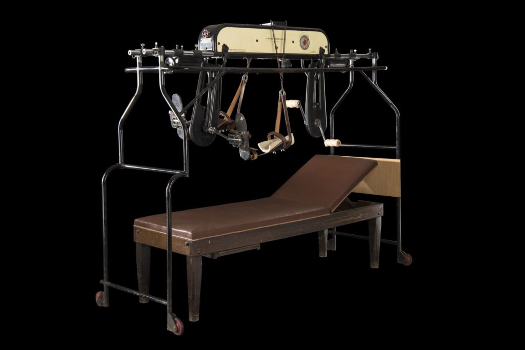
On the first day of the 1948 Summer Olympics in London, sixteen ex-servicemen took to the lawn of the Stoke Mandeville Hospital in Aylesbury to compete in the first ever Stoke Mandeville Games. It was organised by Dr Ludwig Guttmann (1899-1980), a Jewish neurosurgeon and refugee from Germany, who set up a specialist Spinal Injuries Unit at the hospital in 1944. From these small beginnings, the event grew into what we now know as the Paralympic Games.
Beds like this were part of a new exercise regime introduced by Guttman to help war veterans with spinal injuries build physical and mental strength. At the time, people with spinal injuries were often seen as beyond recovery – confined to beds, with little hope.
Guttmann wanted his patients to leave hospital as confident and able members of the community, and he saw physical activity as crucial in achieving this. Patient’s using the Bed cycle would push the pedals with their hands, building muscle in their arms and upper body. Once stronger, they were encouraged to use a wheelchair and take part in games such as darts or snooker. Sport soon became an integral part of Stoke Mandeville’s rehabilitation program and remains so today.
STOP 5: Shocking the heart back into rhythm
Continue to walk down the walled showcase to find the display called Electricity and Health. Look for this silver object on the far right hand side of the showcase.
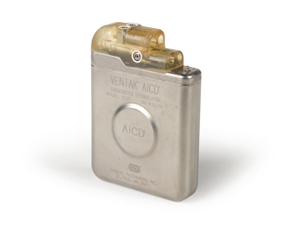
This metallic object is an implantable defibrillator, designed to detect a dangerous heartbeat—and shock the heart back into rhythm.
This life-saving idea came from Mieczysław Mirowski (1924-1990), a Polish-born Jewish doctor, inspired after a colleague died suddenly from an irregular heartbeat in 1968. The first one was implanted in 1980 after years of research.
Early versions were bulky and required major surgery – but they worked. Today, these devices are far smaller and save countless lives worldwide.
Stop 6: How Ernst Chain helped turn Penicillin into a life-saving drug
Walk further along the wall showcases until you arrive at the Penicillin display.
You may have heard of Alexander Fleming, who discovered the penicillin mould in 1928. But you might not know the name Ernst Chain (1906-1979). He was a German born Jewish biochemist. After the Nazis came to power, Chain left Germany to move to England. His mother and sister stayed in Berlin, and he later learned that they died in a concentration camp.
Chain joined a large team of scientists in Oxford in the 1940s investigating microorganisms that produced germ-killing compounds. Chain with his colleague Howard Florey, began to re-examine Flemings work on Penicillin. They went on to discover how the penicillin mould killed bacteria, and crucially how to turn penicillin into a useable medicine.
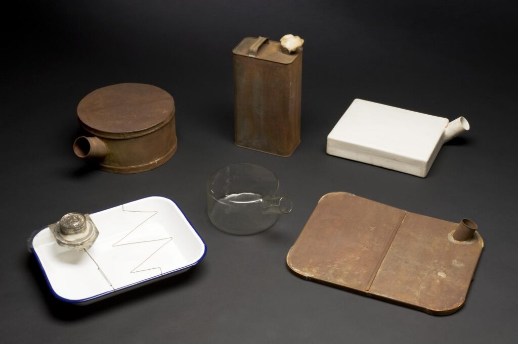
The apparatus on display was used by Chain and Florey, in their earliest experimental work. It was used to grow the mould which produces penicillin, on the surface of a shallow nutrient soup. The containers, once biscuit tins and butter containers, were plugged with cotton wool, to stop air bringing in other microbes that could contaminate the growing process. Drawing on the experience with this apparatus, specially designed ceramic vessels were soon developed. Later, Chain pioneered new techniques to produce penicillin on an industrial scale.
Chain and the Oxford team’s work on producing this ‘wonder drug’ saved millions of lives. For the first time, doctors could reliably treat bacterial infections that had once been deadly. Penicillin marked the beginning of the antibiotic era and earned Chain, Florey and Flemming a Nobel Prize in 1945.
STOP 7: Healing Roots
Walk towards the end of the gallery into the Faith, Hope and Fear Gallery, move past the display of religious healing figures towards the square showcase displaying many charms and amulets.
Can you spot this small piece of iris root amongst the many charms and amulets on display?
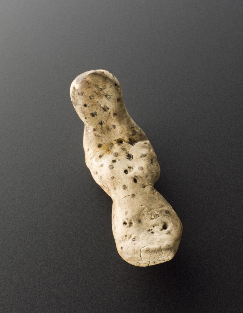
With its vaguely human-like shape, the root was once carried as a protective charm and was believed to have curative properties. It may have been used to reduce the pain of childhood teething as iris root rubbed on the gums was at that time a common way to relieve pain.
The root was acquired in Whitechapel, in the East End of London, in 1900 by Edward Lovett (1852-1933). Lovett was a collector of British amulets and charms and documented different medical traditions and beliefs that he considered were at risk of being lost. He believed that carrying such roots was a tradition associated with Jewish communities living in London’s East End at that time.
Whilst not a technological innovation, for many Jewish families, the use of these kinds of herbal or folk remedies would have been common and have played an important part within Jewish medical history.
A destination for many different immigrants and refugees, including those from different Jewish communities, London’s East End brought together many different cultural, medical and folk traditions over the centuries.
STOP 8: Bringing Treatment Home: Milton Roy Kidney Dialysis Machine c.1966
Turn left and walk to the third showcase, where you will find this kidney dialysis machine.
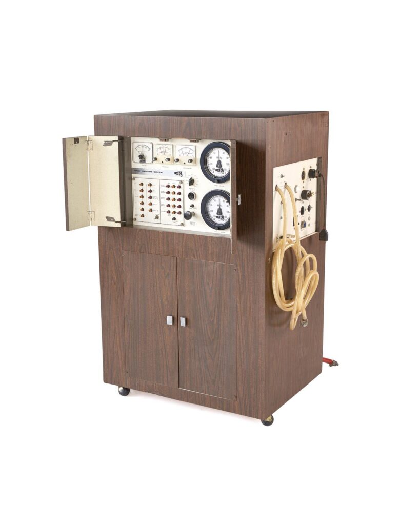
This dialysis machine once stood in the home of Moreen Lewis. It was used to remove waste from her blood after her kidneys failed between 1966 until her death in 1976. The machine’s wooden veneer was designed to make it look like other household furniture to fit in for use at home.
Moreen was one of the first patients in the UK to be treated by Stanley Shaldon, a London-born doctor from an Orthodox Sephardi Jewish family. His family made the decision to change their surname from Schlaff to Shaldon in 1943, as a consequence of rising antisemitism and xenophobia during the Second World War.
Shaldon pioneered new ways to treat patients with kidney disease to give them more control over their treatment.
In the 1960s, dialysis was rare. Machines were expensive and based in hospitals. Many patients were not able to access treatment. Those lucky enough to be treated found they spent much of their lives in hospital as dialysis took many hours several times a week.
Shaldon wanted to change how dialysis impacted the lives of patients like Moreen. In 1966 he set up the National Kidney Centre in an ordinary house in London to treat and train patients and their families in the techniques of carrying out dialysis at home, overnight, with machines such as this.
’What struck me as being very interesting was the concept of self-treatment‘, Shaldon said. ’That you could inspire in somebody who had a potentially fatal disease…a sense of independence, not dependence.’
This self-guided trail of objects in the Science Museum has been published for Jewish Cultural Month, which is taking place in the UK from 16 May – 16 June 2026.