During the middle decade of the nineteenth century, the inner workings of the human eye were explored for the first time thanks to the invention of the ophthalmoscope. The Atlas of Ophthalmoscopy by Richard Liebreich offered the medical profession the means to understand a living retina.
Liebreich’s book simultaneously championed and harnessed the technological development of the ophthalmoscope, while also offering a brilliant guidebook for the identification and treatment of fundus disease. A third edition of this 1863 publication is on display in the Science Museum’s Glimpses of Medical History gallery.
Richard Liebreich, who was born in Konigsberg, Germany in 1830, was able to work with some of the main protagonists in the development of modern ophthalmology. The second half of the nineteenth century saw significant changes in how ophthalmological medicine was understood, with many of these changes taking place in Germany. As a consequence of this, Germany is still regarded as a centre of ophthalmological research excellence to this day.

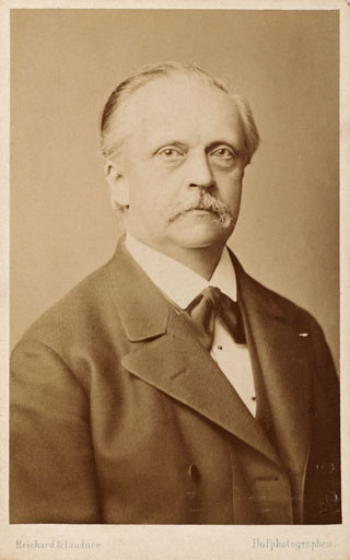
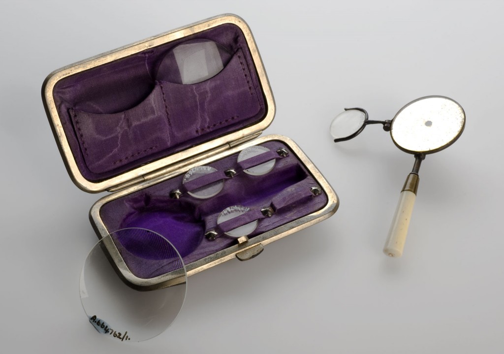
Liebreich worked as Hermann Helmholtz’s assistant, who in 1851 invented the first ophthalmoscope capable of viewing the internal workings of human eyes. British engineer and mathematician, Charles Babbage is recognised in some quarters for inventing the ophthalmoscope in 1847.
However, his failure to promote the discovery ensured that the majority of credit was passed to Helmholtz. Liebreich also worked as the assistant of famed ophthalmologist Albrecht von Graefe between the years of 1854 and 1862, and during this time also devised his own ophthalmoscope that improved upon Helmholtz’s original design.

Liebreich’s Atlas of Ophthalmoscopy was dedicated to Helmholtz and Von Graefe, and contained 57 colour drawings of the eye. Liebreich benefitted from being an incredibly skilled artist, and it was this ability which underpinned the success of his Atlas. The founder of the Royal Eye Hospital in London, John Zachariah Laurence, described the images contained within the Atlas as “scrupulous copies of nature”.

The great quality of the Atlas was that it mapped and recorded the inner workings of the eye, successfully combining technological improvements with medical understanding. The Atlas gave doctors around the world beautifully comprehensive comparisons between healthy and diseased retinas, as well as demonstrating the appearance of certain optical conditions.
Liebreich’s Atlas vividly depicted the inner eye nearly twenty years before accurate photography was possible, and as such made significant contributions to the burgeoning discipline of scientific ophthalmology.
The Atlas had worldwide reach and influence. It was originally published simultaneously in French and German, but soon after versions appeared in both Spanish and English. The third edition currently on display in the museum is from 1885.
Liebreich’s work ensured that he was a respected figure within nineteenth century ophthalmology and in 1870 he was offered a significant role at the newly opened St Thomas’s Hospital in London.
The Lancet opposed his appointment because they felt it was “a gratuitous and unwarrantable insult to English ophthalmologists”. The Medical Times and Gazette defended Liebreich, insisting that his close associations with the German school were to be of huge importance to the fledgling hospital. They reasoned that in order to advance St. Thomas’s, and English ophthalmology in general, they had to “begin by assimilating all that Germany” (and hence Liebreich) had to teach.
Upon leaving his post at St Thomas’s Hospital in 1878 Liebreich slowly withdrew himself from the influential central spheres of ophthalmological medicine, focusing his energies instead on the impact optical disease had on the paintings of artists such as J.M.W Turner. He died in Sicily in 1917 having contributed significantly to the previous centuries advancements in the study of ophthalmology.
2 comments on “Richard Liebreich’s Atlas of Ophthalmoscopy”
Comments are closed.
Atlases such as this were important for the medical profession not just because they showed retinal changes in eye disease but could be used to map stages in vascular disease in a living person.
In addition they could also be used by non-medically trained people to see how dis-ease affected living tissue. In particular the new breed of nineteenth century sight-testing opticians could use this type of atlas combined with the use of ophthalmoscopes to distinguish between mechanical visual problems which could be best treated with spectacles and medical visual problems which needed to be referred to medical professionals.
Thank you for your reply Sandra,
As you point out, Atlases such as these were important tools for distinguishing between various medical issues, and they were significant because they visually mapped parts of the body and stages of disease. Atlases added a layer of knowledge and understanding to medical equipment, and ensured that what was being seen could actually be interpreted for the benefit of the medical profession at large. Communicating how to use and understand a newly discovered medical tool is a significant aspect of medicines overall development.
In the blog I aimed to show the increased dialogue which existed in the ophthalmic profession during the nineteenth century and the growing confidence that people like Liebreich had for explaining and recording a “new” sphere of the human body. The tight-knit German school of individuals who Liebriech worked with were fundamental in expanding the understanding and knowledge of their profession and for encouraging an increasingly scientific dialogue to surround and support their work. For example, in 1854 Von Graefe started the journal Archiv fur Ophthalmogie, a periodical which continues to this day, under the new title of “Graefe’s Archive for Clinical and Experimental Ophthalmology”.
Thanks very much,
Jack