The 1953 discovery of the double helix molecular structure of DNA, the long chain-like molecule deoxyribonucleic acid, is a key moment in modern history.
DNA, the molecule in our cells that carries information in the form of genes, was revealed to be a twisting pair of strands that has been unravelling to replicate itself since the dawn of life some four billion years ago.
The find revealed the molecular details of how traits are passed down generations and the seminal paper on this milestone in molecular biology by James Watson and Francis Crick in Cambridge was published in the journal Nature 70 years ago this week.
However, today in the journal Nature, new evidence is published that shows Rosalind Franklin was truly an equal contributor in the discovery of DNA’s structure and, moreover, Watson and Crick were not as caddish as they are often portrayed, in a commentary by Matthew Cobb of the University of Manchester and Nathaniel Comfort of Johns Hopkins University, Baltimore.
It is also more than half a century since Watson wrote The Double Helix, his notoriously candid and raffish first-hand account of what he called ‘perhaps the most famous event in biology since Darwin’s book’.
By popular accounts, the ‘eureka moment’ in the discovery of the double helix came when Watson was shown an X-ray image of DNA taken by Franklin’s student, Raymond Gosling, without her permission or knowledge.
Known as Photograph 51, this image is treated as ‘the philosopher’s stone of molecular biology’, write Cobb and Comfort. When it was ‘stolen’ by Watson and Crick, ‘it became the emblem of both Franklin’s achievement and her mistreatment,’ they explain.
In this traditional version of events, Franklin is portrayed as brilliant but ultimately unable to decipher exactly what her own data were telling her about DNA, when Watson supposedly understood the significance of Photograph 51 at first glance.
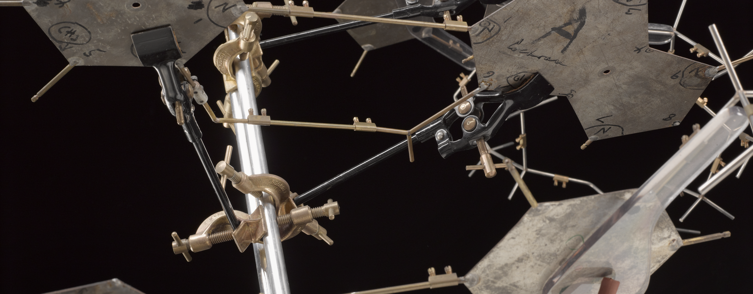
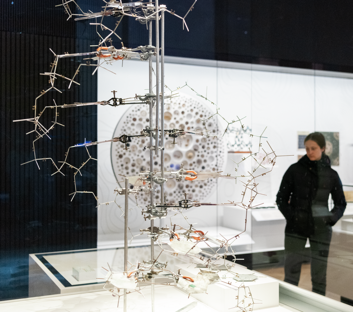
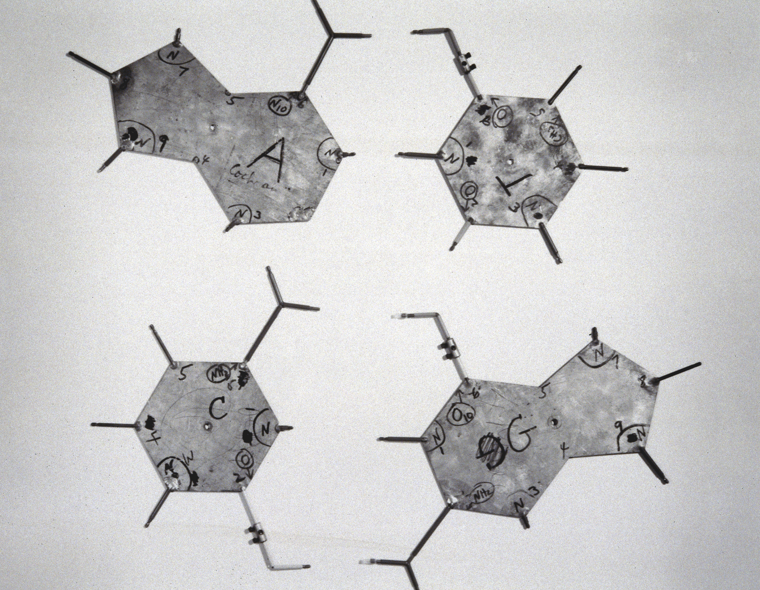
The resulting metal model of DNA that he and Crick put together after experimenting with cardboard cut-outs, and which was used in a famous Time magazine picture, can be seen in Medicine: The Wellcome Galleries in the museum.
On visiting the Churchill College archives in Cambridge, the authors found a hitherto unstudied draft news article — written by journalist Joan Bruce in consultation with Franklin and meant for publication in Time magazine — and later found an overlooked letter from one of Franklin’s colleagues to Crick in the Crick archive at the University of California, San Diego.
This new evidence suggests Franklin did not fail to understand the structure of DNA. Cobb and Comfort argue that Franklin, along with Maurice Wilkins at a Medical Research Council unit in King’s College London, was ‘one half of the team that articulated the scientific question, took important early steps towards a solution, provided crucial data and verified the result’.
One complication neglected in popular accounts is that there were two kinds of DNA under study with X-rays at King’s: Franklin focused on the crystalline A form, consisting of DNA alone, while Wilkins focused on a wet B form of DNA.
The X-ray scattering patterns of the latter were easier to interpret and suggested a helix, even to Franklin. But she was more interested in the A form of pure DNA which yielded complex X ray diffraction images that were much harder to understand. That is why in late 1952 and early 1953, she rejected the idea that DNA was a helix, even writing a mock funeral notice for the helix, believing that the X ray patterns created by the B form were an artefact of how the DNA was waterlogged.
As Cobb and Comfort write, ‘Her focus on the drier A form ignored the very wet reality of the inside of a cell — which would mean that DNA took the more humid B form. Together with her insistence that the diffraction data be fully analysed before any modelling was attempted, it would hamper Franklin’s efforts for more than a year.’
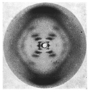
There is far too much emphasis on Photograph 51 in traditional accounts, they add. ‘Various lines of evidence — including The Double Helix itself, read carefully — show that it played little, if any, part in Watson and Crick’s inching towards the correct structure between January and March 1953. In fact, it was other data from Franklin and Wilkins that proved crucial, and even then, what really happened was much less malicious than is widely assumed.’
Shortly after Watson saw Photograph 51, Crick’s supervisor, Max Perutz, handed him and Watson an informal report about the activity of the King’s MRC unit, which he had been given as part of an official visit to the unit in December 1952, which describes Franklin’s work. The letter suggests that, indeed, Watson and Crick had seen key details of the B form she had deduced and, no, they had not stolen her data, as often claimed: Perutz had handed them over.
The gist of what really happened in 1953 is also reflected in Bruce’s unpublished article, which portrayed the work as being done by ‘two teams’: one, consisting of Wilkins and Franklin, gathering experimental evidence using X-ray analysis; ‘the other’ comprising Watson and Crick, working on theory.
This was further underlined by one of the first public presentations of the double helix, at the Royal Society Summer Exhibition of June 1953 (then called a Conversazione), where the discovery was shown as the work of both the King’s and the Cambridge groups, with Franklin as the lead author.
‘At the time,’ said Cobb, ‘this was seen as collective work, partly because the stakes were pretty low – the evidence for the genetic role of DNA in all life was still unclear (and remained so for many years), although the structure, when revealed, strongly suggested that it was.’
In conclusion, Franklin has been reduced to the ‘wronged heroine’ of the double helix by traditional accounts when she should be remembered as ‘an equal contributor’ to the solution of the structure, they argue: sadly, her role could not be acknowledged in the Nobel prize shared in 1962 because she died of ovarian cancer at the age of 37 in 1958.
Her story has many modern resonances, add Cobb and Comfort: ‘She was up against not just the routine sexism of the day, but also more subtle forms embedded in science — some of which are still present today.’
Fittingly, Nature reveals that the order of authors of the new paper was determined ‘by the toss of a Rosalind Franklin 50p coin.’