Galleries you will visit: Medicine: The Wellcome Galleries, Making the Modern World, Mathematics: The Winton Gallery and Wonderlab
Stop 1: Louis Pasteur’s compound microscope
Make your way to Level 1 where you will find Medicine: The Wellcome Galleries. Here, you will find Louis Pasteur’s compound microscope.
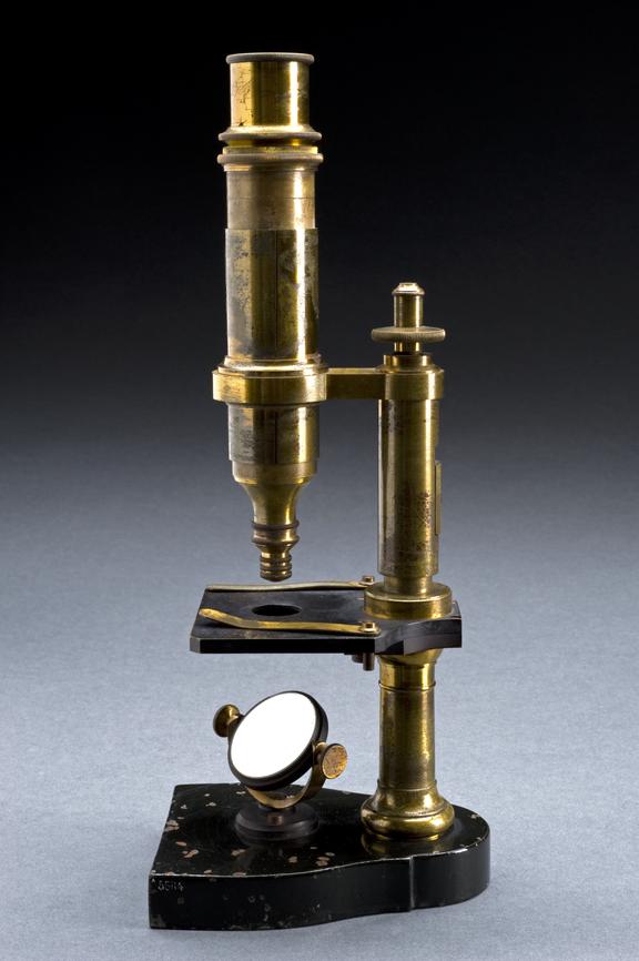
Chemist and microbiologist, Louis Pasteur, used microscopes like this during his experiments on spontaneous generation – the theory that living creatures could arise from nonliving matter and that such processes were commonplace and regular.
By 1864, Pasteur disproved this theory by experimenting with fermentation. He placed yeast water in a swan-necked flask that only allowed air to enter. The water remained clear. Only when the flask was open to dust and micro-organisms did fermentation occur.
Stop 2: Crystal model of magnesium ammonium phosphate
Also found in Medicine: The Wellcome Galleries is this crystal model of magnesium ammonium phosphate (Mg NH4 PO4 6H2O).
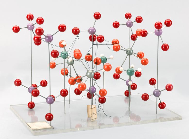
This model is from a collection relating to the X-ray crystallography research associated with Dame Kathleen Lonsdale’s work in investigating the cause of kidney stones.
Stop 2: Density map of a myoglobin molecule
Make you way up to Level 2 and enter the Mathematics: The Winton Gallery. Here, you’ll find the electron density map of a myoglobin molecule.

Myoglobin (symbol Mb or MB) is an iron- and oxygen-binding protein found in muscle tissue of vertebrates (animals with a spinal cord) in general and in almost all mammals. It was the first protein to have its three-dimensional structure revealed by X-ray crystallography.
Stop 3: Dorothy Hodgkin’s model showing the structure of insulin
Using Lift D, head back down to Level 0. Once you are out of the lift, turn right and enter Making the Modern World.
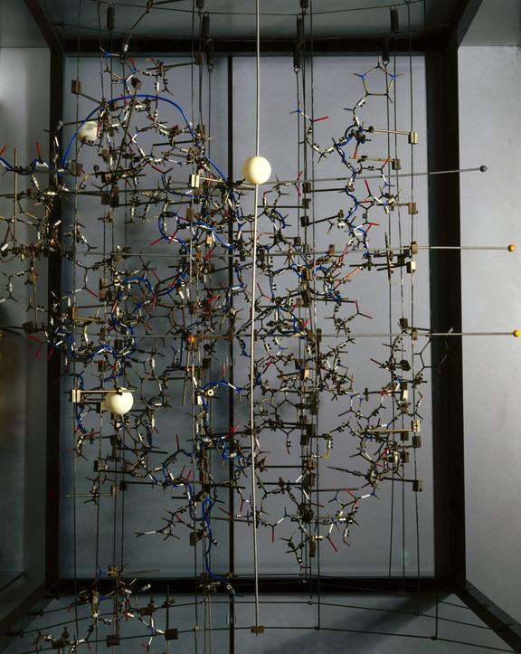
In this gallery, you will find British chemist and crystallographer Dorothy Hodgkin’s model showing the structure of insulin, a hormone produced by the pancreas to break down sugars in the body.
In 1935, Dorothy M Crowfoot Hodgkin published the first X-ray photograph of insulin. However, Hodgkin and her team were unable to determine the 3-D structure of insulin until 1969, when this model was made. The larger metal balls in the model represent zinc, which was introduced chemically into the protein to decode the rest.
Stop 4: Oscillation-rotation x-ray diffraction camera
While you’re in Making the Modern World, look out for the Oscillation-rotation x-ray diffraction camera used by physicist Professor John Desmond Bernal.
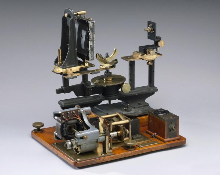
Bernal used this X-ray diffraction camera at the Royal Institution in London.
When X-rays are passed through crystals they scatter to create a pattern that can be used to determine the structures of molecules.
Known today as X-ray crystallography, it was a crucial technique used to understand the structure of penicillin, DNA and insulin. Bernal was also interested in the social function of science and wrote widely on the history of science.
Stop 5: Wonderlab
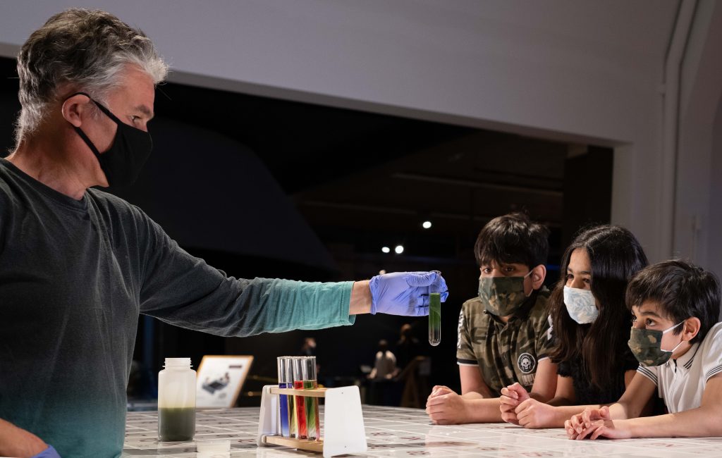
And last, but certainly not least, it’s time to get hands-on with chemistry.
Make sure to visit the Chemistry Bar in Wonderlab on Level 3, where our team of fabulous science Explainers will conduct live science demos.
Our fully interactive Wonderlab has over 50 hands-on exhibits and will inspire you to see the world around you in new and exciting ways.
Where to eat:
On Level 0 you will find the Energy Cafe if you fancy treating yourself to lunch, or with one of our homemade cakes and an award-winning coffee.
The Science Museum is every day from 10.00–18.00 (last entry 17.15). Head to our website to pre-book your free tickets.
Want more? Delve into our online stories of how experimentation and innovation in chemistry affects the world around us.